Quiz · Medium
Core calculations and concepts across the major imaging modalities, from the X-ray tube to PET.
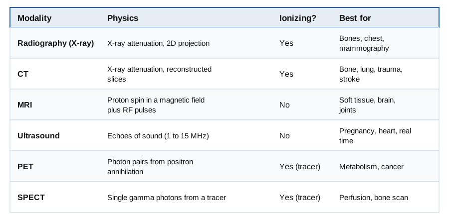
Which of these imaging modalities does NOT use ionizing radiation?
CT
PET
MRI
Radiography
MRI uses a static magnetic field and radiofrequency pulses. CT and radiography use X-rays; PET uses gamma photons from a radiotracer.
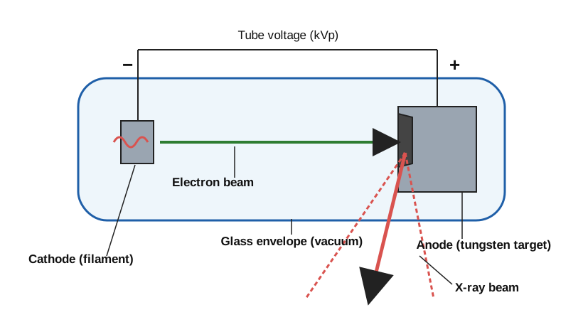
In an X-ray tube, increasing the tube current (mA) mainly increases:
The number of photons produced
Maximum photon energy
The target's atomic number
The wavelength of characteristic lines
mA sets the electron count and thus photon quantity. kVp sets the maximum photon energy, and the characteristic lines depend only on the target material.
What is the maximum photon energy produced at 90 kVp?
30 keV
45 keV
180 keV
90 keV
An electron accelerated through 90 kV can give all its energy to one photon, so the maximum is 90 keV. The mean is lower (about one third to one half).
Roughly what fraction of the electron energy in a diagnostic tube becomes X-rays?
20%
1%
50%
99%
About 99% appears as heat, which is why anode heat capacity limits exposures.
Which interaction gives the best bone-to-soft-tissue contrast at diagnostic energies?
Compton scattering
Pair production
Coherent scattering only
Photoelectric absorption
Photoelectric probability varies as Z³/E³, so bone (calcium) absorbs much more than soft tissue. Compton depends on electron density, which is similar in both.
If μ = 0.1 cm⁻¹, what is the half-value layer?
6.93 cm
0.69 cm
10 cm
1 cm
HVL = 0.693/μ = 0.693/0.1 = 6.93 cm. 0.69 cm results from using μ = 1.
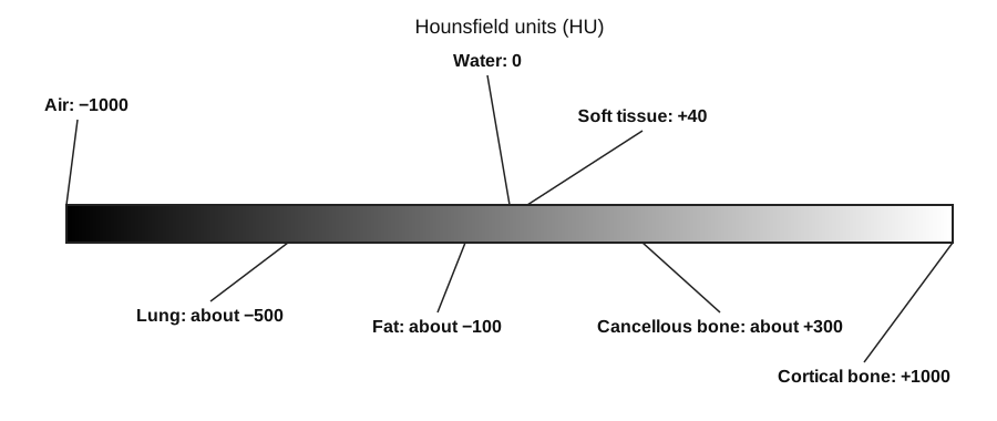
On the Hounsfield scale shown, water has a CT number of:
-1000
+40
0
+1000
Water is defined as 0 HU and air as -1000 HU. Soft tissue is about +40.
Which tissue has the lowest CT number?
Water
Fat
Muscle
Cortical bone
Fat is about -100 HU, below water (0), muscle (+40) and bone (hundreds). Only air and lung are lower than fat.
CT reconstruction by filtered back projection applies which filter to projections?
Low-pass
Notch
Ramp (high-pass)
Median
The ramp filter compensates for the 1/r blur from simple back projection.
Dark streaks between two dense objects (dental fillings) in CT are due to:
Beam hardening and photon starvation
Ring artifact
Motion
Partial volume
Dense metal hardens and starves the beam, producing streaks. Rings come from detector problems.
A source reads 16 mGy/h at 1 m. The dose rate at 4 m is:
8 mGy/h
4 mGy/h
0.25 mGy/h
1 mGy/h
Inverse square law: 16/4² = 1 mGy/h. Using 1/r gives the wrong 4.
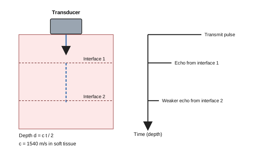
In the pulse-echo diagram, the time between transmit and the second echo is proportional to:
The frequency
The depth of interface 2
The speed of the transducer
The amplitude
Round-trip time t = 2d/c, so t is proportional to depth.
An echo returns 65 µs after transmission in soft tissue. The reflector depth is about:
10 cm
1 cm
20 cm
5 cm
d = ct/2 = 1540 × 65×10⁻⁶/2 ≈ 0.050 m = 5 cm (the 13 µs per cm rule). 10 cm results from forgetting the factor of 2.
Which change improves axial resolution in ultrasound?
Higher frequency / shorter pulses
Lower frequency
Longer pulses
Smaller transducer
Axial resolution is SPL/2, so shorter pulses and higher frequency are better, but penetration falls.
Why is gel needed between transducer and skin?
To cool the transducer
To increase frequency
To remove air which would reflect almost all the sound
To polarize tissue
The large impedance mismatch of air and skin reflects more than 99% of the beam.
The Doppler shift is zero when the beam angle to flow is:
0°
90°
30°
60°
Δf ∝ cosθ and cos 90° = 0.
The Larmor frequency of hydrogen at 3 T is about:
21 MHz
64 MHz
128 MHz
300 MHz
f = 42.58 MHz/T × 3 T ≈ 128 MHz. 64 MHz is the value at 1.5 T.
A T1-weighted image uses:
Short TR, short TE
Long TR, long TE
Long TR, short TE
Short TR, long TE
T1 weighting needs a short TR and short TE. Long TR and long TE gives T2 weighting.
On a T2-weighted image, CSF appears:
Dark
Isointense with fat
Invisible
Bright
Fluid has a long T2 so it stays bright; on T1 it is dark.
Gadolinium contrast agents enhance tissue on T1 images because they:
Lengthen T1
Shorten T1
Block RF
Increase B0
Gadolinium is paramagnetic and shortens T1, so enhancing tissue recovers quickly and is bright.
PET detects:
A single 140 keV photon
Radio waves
Ultrasound echoes
Pairs of 511 keV photons in coincidence
Positron annihilation yields two 511 keV photons at about 180 degrees, detected in coincidence by a detector ring.
True or false: SPECT usually has better spatial resolution than PET.
True
False
SPECT is around 10 mm, PET about 4-6 mm, because PET uses coincidence detection rather than lead collimators.
A radiotracer has a half-life of 6 hours. After 18 hours what fraction remains?
12.5%
50%
25%
6.25%
18 hours is three half-lives: (1/2)³ = 12.5%.
If dose is increased by a factor of 4 (quantum noise limited), SNR increases by:
4
8
2
16
Quantum noise is Poisson, so SNR ∝ √N and quadrupling N doubles SNR.
