Quiz · Medium
Core neuroanatomy from the lobes and subcortical structures to the meninges, CSF, spinal cord tracts and cranial nerves.
Which lobe contains the primary visual cortex?
Occipital lobe
Frontal lobe
Temporal lobe
Parietal lobe
V1 sits at the occipital pole. Visual association areas extend into the temporal and parietal lobes, but the primary cortex is occipital.
The central sulcus separates which two lobes?
Parietal and occipital
Frontal and temporal
Temporal and parietal
Frontal and parietal
The motor strip lies in front of it and the somatosensory strip behind it. The lateral sulcus separates the temporal lobe from the frontal and parietal lobes.
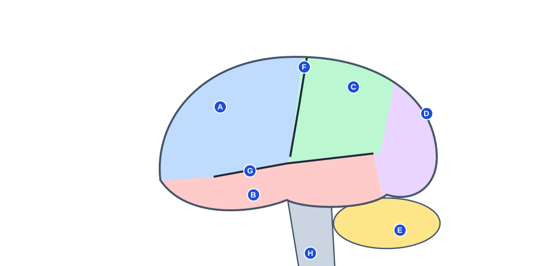
Which structure is marked B in the diagram?
Frontal lobe
Parietal lobe
Temporal lobe
Occipital lobe
The temporal lobe lies below the lateral sulcus and handles hearing, language comprehension and memory.
Which structure is marked F in the diagram?
Parieto-occipital sulcus
Central sulcus
Longitudinal fissure
Lateral sulcus
The central sulcus runs between the precentral (motor) and postcentral (somatosensory) gyri.
Which structure is marked C in the diagram?
Frontal lobe
Parietal lobe
Occipital lobe
Temporal lobe
The parietal lobe lies behind the central sulcus.
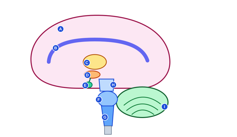
Which structure is marked B in the diagram?
Corpus callosum
Pons
Fornix
Thalamus
The corpus callosum is the arched white-matter band linking the two hemispheres. The thalamus sits beneath it.
Which structure is marked D in the diagram?
Thalamus
Pituitary gland
Midbrain
Hypothalamus
The hypothalamus sits below the thalamus and above the pituitary, which it controls.
Which structure is marked F in the diagram?
Cerebellum
Midbrain
Pons
Medulla oblongata
The pons is the bulge of the brainstem between the midbrain and medulla, in front of the cerebellum.
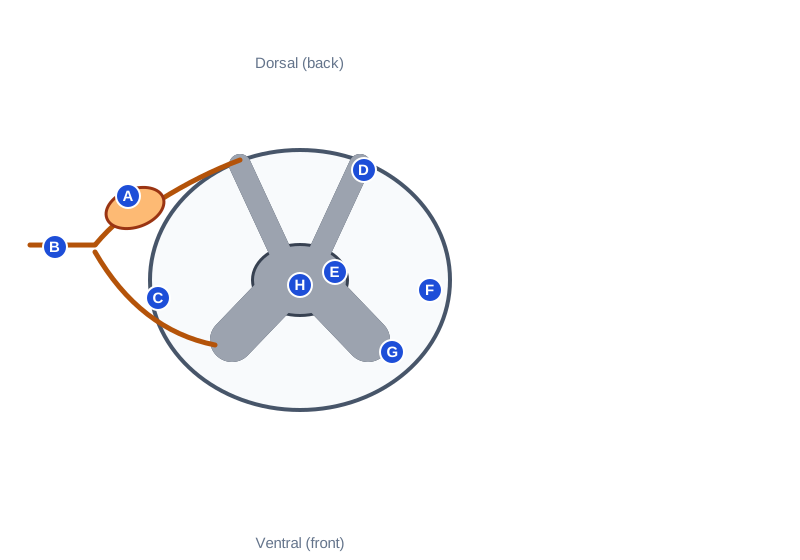
Which structure is marked G in the diagram?
Central canal
Dorsal horn
Ventral horn
Dorsal root ganglion
The ventral horn holds the cell bodies of lower motor neurons. The dorsal horn receives sensory input.
Which structure is marked A in the diagram?
Ventral root
Dorsal root ganglion
Spinal nerve
Central canal
The dorsal root ganglion contains the cell bodies of sensory neurons.
A patient cannot form new memories after bilateral damage to the medial temporal lobes. Which structure is most likely involved?
Hippocampus
Cerebellum
Amygdala
Thalamus
The hippocampus is essential for forming new declarative memories (patient H.M.). The amygdala is more about emotional learning, such as fear.
True or false: the spinal cord extends to the bottom of the vertebral column in adults.
True
False
The adult cord ends at about L1-L2; the cauda equina continues below, which is why lumbar punctures are safe at L3-L4.
Damage to Broca's area typically causes what?
Fluent speech with poor comprehension
Inability to repeat words only
Complete loss of hearing
Nonfluent speech with relatively preserved comprehension
Broca's area (left inferior frontal gyrus) is for speech production. Fluent but meaningless speech is Wernicke's aphasia; poor repetition alone is conduction aphasia.
Which statement about the corpus callosum is true?
It is part of the brainstem
It produces cerebrospinal fluid
It connects the cerebrum to the cerebellum
It connects the two cerebral hemispheres
It is the major commissure. Choroid plexus produces CSF; the cerebellar peduncles link cerebellum and brainstem.
Which brain region is the main sensory relay station for all senses except smell?
Basal ganglia
Medulla
Thalamus
Hypothalamus
Sensory signals relay through thalamic nuclei (LGN for vision, MGN for hearing, VPL/VPM for touch). The hypothalamus is for homeostasis.
Which part of the brain regulates body temperature, hunger and thirst?
Cerebellum
Hypothalamus
Pons
Thalamus
The hypothalamus maintains homeostasis and controls the pituitary. The thalamus relays sensory input.
Loss of dopamine neurons in the substantia nigra is characteristic of which disorder?
Parkinson's disease
Alzheimer's disease
Multiple sclerosis
Huntington's disease
Parkinson's affects nigrostriatal dopamine neurons. Huntington's involves the striatum; Alzheimer's begins in the medial temporal lobe; MS is demyelination.
A person has an intention tremor and a staggering gait. Which region is most likely damaged?
Cerebellum
Occipital lobe
Primary motor cortex
Hypothalamus
Cerebellar damage causes ataxia, dysmetria and intention tremor. Motor cortex damage causes weakness or paralysis instead.
What is carried by the dorsal root of a spinal nerve?
Only autonomic motor signals
Both sensory and motor equally
Motor (efferent) commands
Sensory (afferent) information
Bell-Magendie law: dorsal = afferent, ventral = efferent. The spinal nerve itself is mixed.
Where do most corticospinal tract fibers cross the midline?
Anterior commissure of the cerebrum
Spinal dorsal horn
Pyramids of the medulla
Midbrain tegmentum
About 85 to 90% decussate in the medullary pyramids, so each motor cortex controls the opposite side.
Which cranial nerve controls the muscles of mastication and carries facial sensation?
Facial (VII)
Trigeminal (V)
Glossopharyngeal (IX)
Hypoglossal (XII)
V3 supplies the muscles of chewing. The facial nerve controls facial expression.
Which cranial nerve innervates the lateral rectus?
Oculomotor (III)
Abducens (VI)
Trochlear (IV)
Trigeminal (V)
LR6 SO4 R3: lateral rectus is VI, superior oblique is IV, the rest are III.
Which are the meninges, from outermost to innermost?
Dura, arachnoid, pia
Dura, pia, arachnoid
Arachnoid, dura, pia
Pia, arachnoid, dura
CSF flows in the subarachnoid space between the arachnoid and the pia.
Where is most cerebrospinal fluid produced?
Arachnoid granulations
Pia mater
Dorsal root ganglia
Choroid plexus in the ventricles
Choroid plexus secretes CSF. Arachnoid granulations absorb it back into venous blood.
Which is the correct path of CSF flow?
Third ventricle, lateral ventricles, aqueduct, subarachnoid space
Fourth ventricle, aqueduct, lateral ventricles, third ventricle
Lateral ventricles, third ventricle, cerebral aqueduct, fourth ventricle, subarachnoid space
Lateral ventricles, fourth ventricle, cerebral aqueduct, third ventricle
The aqueduct of Sylvius connects the third and fourth ventricles; blockage there causes obstructive hydrocephalus.
True or false: the right cerebellar hemisphere controls the left side of the body.
False
True
The cerebellum acts ipsilaterally (its pathways cross twice), unlike the cerebral cortex, which controls the opposite side.
